Celebrating SIU’s Saluki TRADITION, Saluki PRIDE and Saluki PROMISE for SIU’s 150thAnniversary, This Is SIU is publishing a monthly feature detailing the past, present and future of notable places, events and people on campus.
They come with names longer than a city block: the laser-induced breakdown spectroscopy, or the Fourier-transform infrared spectrometer. Those, in turn, lead to a parade of acronyms, like NMR and AFM.
They are highly technical, phenomenally revelatory pieces of machinery, pulling back the veil of the unknown for scientists for more than half a century. They are the advanced, top-of-the-line scientific instruments that scientists and researchers use to probe the truths invisible to the naked eye.
And SIU has always been a leading repository for the highest of the high-tech.
There are millions of dollars-worth of such machinery stashed around campus, setting SIU apart from so many others as a major research university.
“SIU Carbondale has been incredibly successful in bringing in funding for the purchase of important research equipment over the last two decades,” said Gary Kinsel, professor and interim vice chancellor of research at SIU. “Our success in this area speaks directly to the collaborative nature of our research faculty and the shared goal of maintaining our status as a High-Research-Activity Doctoral Research University.”
Millions of dollars in machines
The university’s treasure trove of scientific instrument is scattered about the campus, from far-flung McLafferty Annex to the Neckers science building, to the Engineering building, Agricultural Sciences building and life science buildings, as well a few other places. It’s housed in places such as the Mass Spectrometry Facility and the Nuclear Magnetic Resonance Facility, both university-wide assets with instruments available for use by SIU researchers.
To a large extent, the National Science Foundation Major Research Instrumentation Program has paid for the equipment. Over the years SIU brought in more than $2.5 million dollars of research equipment, including a mass spectrometer for proteomics, a solid-state probe for nuclear magnetic resonance spectroscopy, a field-emission scanning electron microscope and an isotope ratio mass spectrometer, Kinsel said. Most recently the university received funding to purchase an atomic force microscope.
But one of the impressive elements of SIU’s success with the program is the required collaborative nature of the grants, Kinsel said.
“This funding program requires that a group of faculty get together and explicitly describe how the presence of piece of equipment on campus will advance their research,” he said. “There are typically five to seven major users, often from many different departments, and another two to six minor users of any piece of instrumentation.”
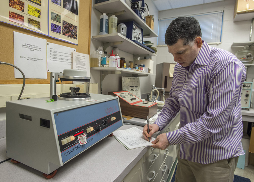
Faculty at a doctoral research university such as SIU are expected to compete for external funding to support their various research activities. The funds not only pay for the actual research equipment and supplies, but also pay for graduate and undergraduate student researcher salaries, Kinsel said.
Competition is stiff. And the more instruments the university has, the better able it is to attract top faculty, owing the research synergies such a concentration can provide. Top faculty mean more funded research grants, and on the cycle goes.
“For some funding sources, less than 1 in 10 proposals will be awarded,” Kinsel said. “For our faculty to compete, the university must be able to provide them with access to cutting-edge research instrumentation.”
But the investment pays dividends for students, researchers and the university as a whole.
“The return on that investment is multiplied many times over for the university,” Kinsel said. “For many of our faculty, it is access to this instrumentation that allows them to successfully compete for funding and, more importantly, to make new discoveries that bring recognition to SIU Carbondale.”
The IMAGE Center
While the machines dot the campus map, the undesignated center of such equipment might be the Integrated Microscopy and Graphics Expertise Center, or IMAGE Center.
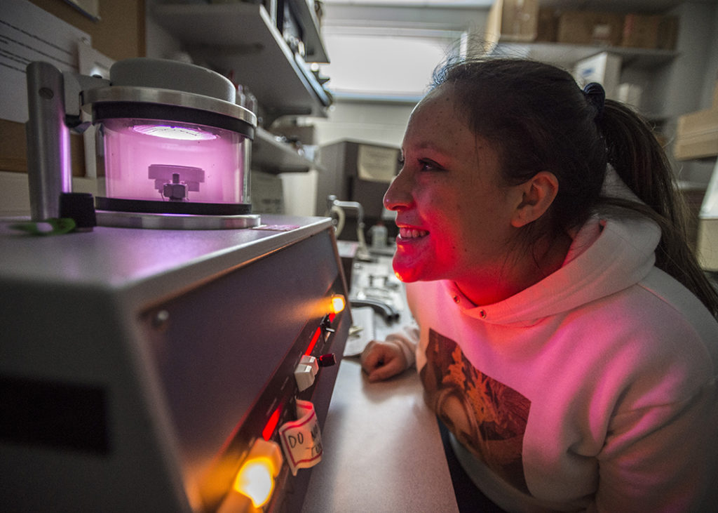
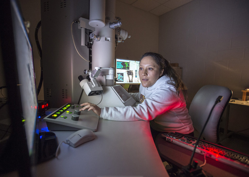
The IMAGE Center provides training, technical service and research assistance with scanning electron, transmission electron, atomic force and light microscopy. Advanced capabilities include X-ray analysis, image analysis and viewing of specimens under near-atmospheric conditions or a vacuum.
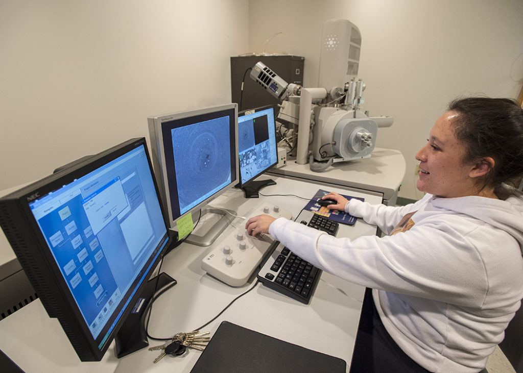
Housed in a small, unobtrusive building just south of Life Science III, the center is home to four of the university’s top machines, including a scanning electron microscope, a transmission electron microscope, fluorescence spectrometer and a soon-to-be upgraded atomic force microscope. It also houses various support equipment used to prepare samples for analysis.
Punit Kohli, professor in the chemistry and biochemistry degree program and director of the IMAGE Center, said the center exists as a resource for all campus researchers, as well as local industry.
“We do our best to be a resource for all faculty, and we work closely with them on their research to provide what services they need,” Kohli said.
Nathalie Becerra-Mora, a doctoral student in chemistry, is one of two graduate students who help keep the IMAGE Center up running. Coming to SIU from Columbia, where she earned her undergraduate degree, was a terrific choice because it has given her wide variety of hands-on experience.
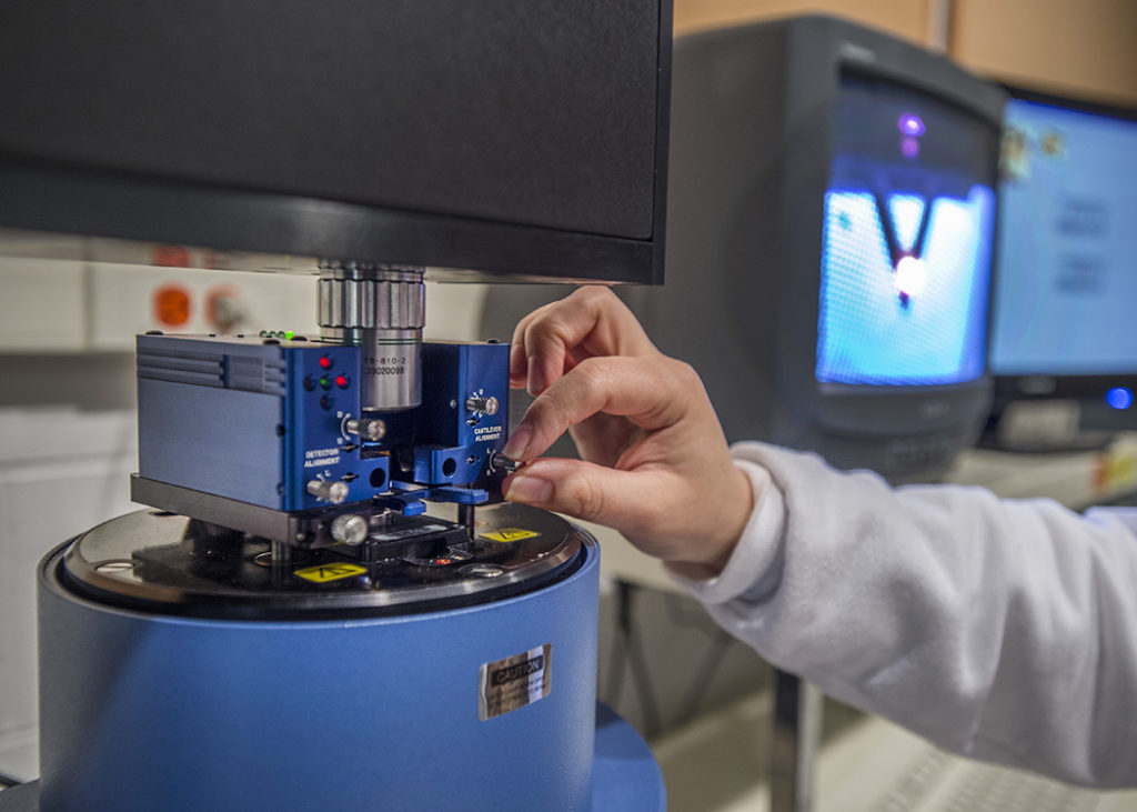
“I’ve had the opportunity to work on so many different instruments and so many researchers on their projects,” Becerra-Mora said. “I would not have received that chance at many other universities.”
Electron microscopy led the way
The specially designed IMAGE Center, which came online in the mid-1990s, is built on a free-floating concrete floor specially crafted to reduce vibration. At such high resolutions and magnifications, even the tiniest bit of shaking can badly reduce the image quality provided by the instruments.
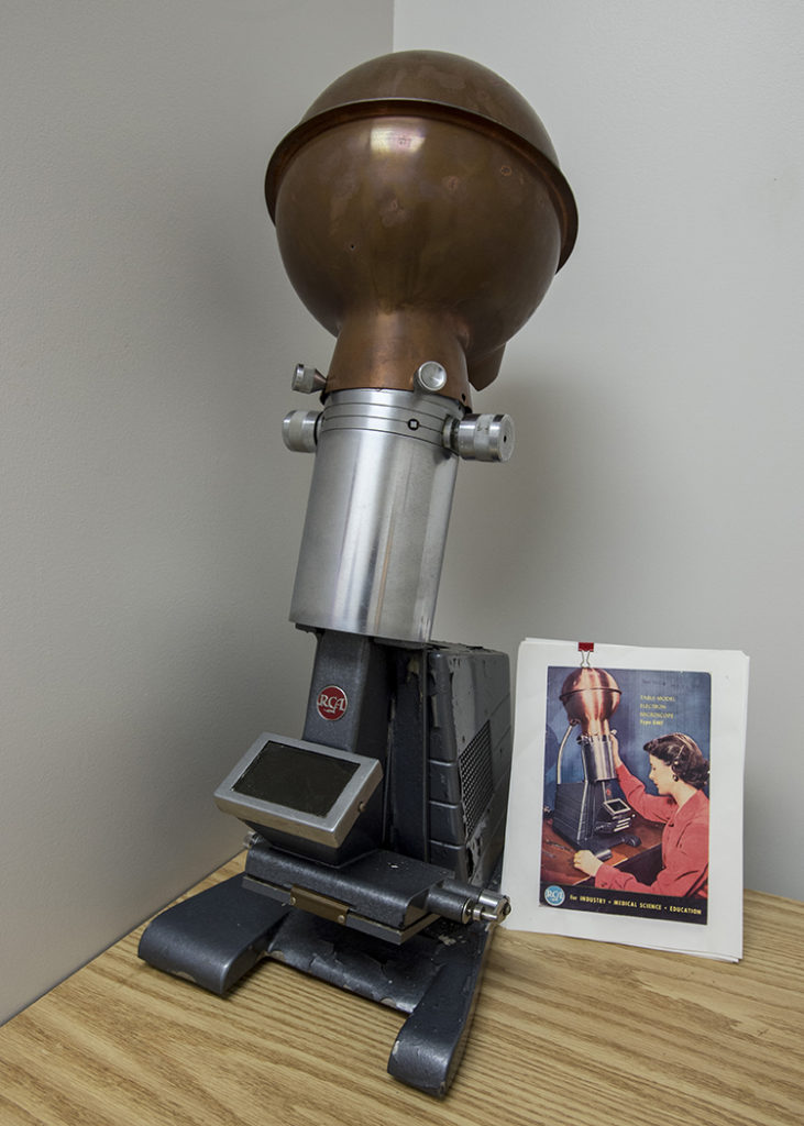
Although the building fairly bristles with modern equipment, two of the most interesting pieces are blast-from-the-past electron microscopes on display. Looking dated, and like something out of a 1950s mad scientist movie, they were once cutting-edge research pieces and are instructive in how far the technology has risen over the decades.
Electron microscopy lets researchers see extremely fine detail biological specimens. When it came on the scene in 1930s, it opened new worlds to the human eye, vastly increasing our knowledge of biological structures and architecturally complex arrangement of macromolecules under the direction of genes. Suddenly, complex structures within cells were clearly seen, as well as biochemical activities associated with each structure. For the first time, too, viruses could be seen and classified.
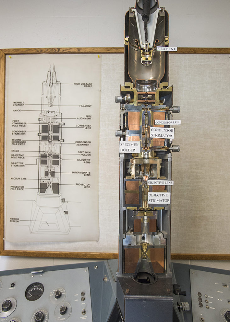
The technology is routinely used in many fields of inquiry including anatomy, anthropology, biochemistry, cell biology, forensic medicine, microbiology, immunology, pathology, physiology, plant biology, toxicology and zoology.
Technology rapidly advances
One now-retired researcher who was in on the ground-floor of electron microscopy at SIU was John Bozzola, who started working with the new technology in the late 1960s as an undergraduate working with an ultramicrotome, a device that takes extremely thin slices of biological material for viewing in a transmission electron microscope. There were two such instruments on campus in those early days, one in the physiology department and the other in microbiology.
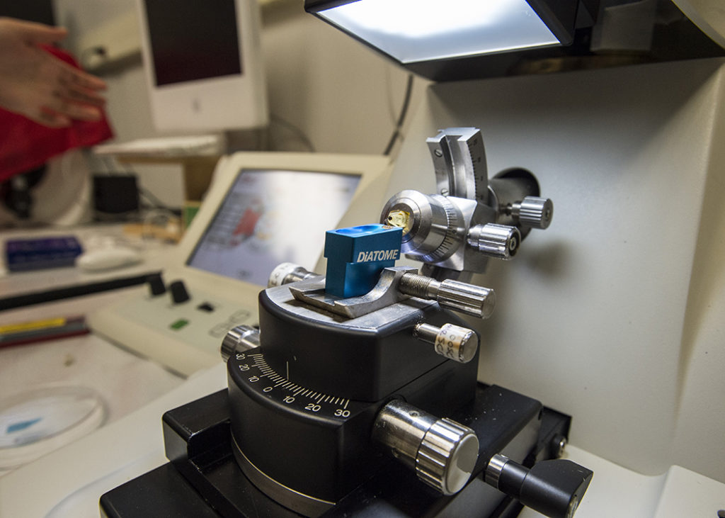
But advances were happening rapidly and soon SIU acquired more advanced machines, he said. During the early 1970s, SIU acquired a Hitachi HU 11AB, which had a considerably better resolution, as well as the ability to rapidly change specimens and films. It was housed at the university’s former animal facility, which the university eventually designated the Center for Electron Microscopy.
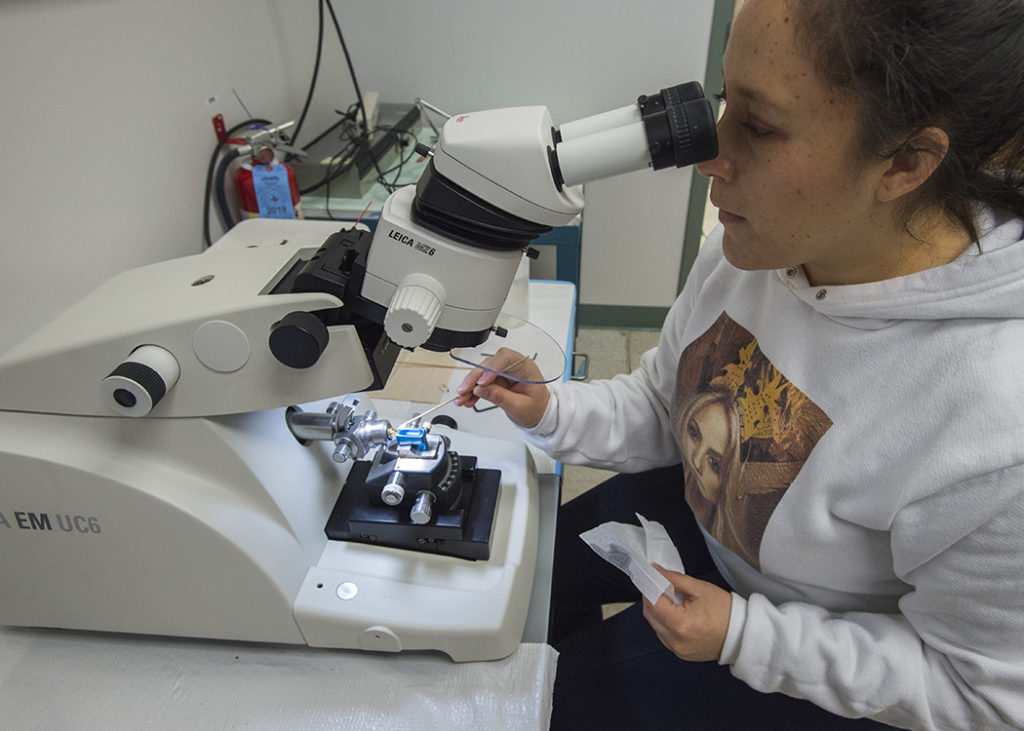
In 1983, Bozzola become the director of the Center for Electron Microscopy, conducting experiments and research and teaching students. Along the way, he oversaw the opening of the CEM’s next evolution: the IMAGE Center, serving as its director starting in 1998. He retired in 2011.
Moving into the new IMAGE Center building 1995, Bozzola said equipment on hand included a new analytical, combined system of a high resolution transmission and scanning electron microscopes, both connected to an X-ray microanalytical system, as well as workhorse electron microscopes from Neckers. They later added an atomic force microscope, one of which is still operating and slated to be replaced in 2020 by a National Science Foundation grant.
University takes cooperative approach
The IMAGE Center may be dedicated solely to such instrumentation. But the machines can be found all over campus, wherever researchers are working.
McLafferty houses such beauties as a hydroacoustics system and the Aquatic Research Laboratory and Saluki Aquarium used for fisheries research, as well as chemistry researcher Sean Moran’s 2D infrared spectrometer, among others. Kohli’s lab at Neckers includes a laser-scanning confocal microscope, a reactive-ion etcher and an ultraviolet-visible absorption spectrometer, while Professor Boyd Goodson’s lab there includes a nuclear magnetic resonance spectrometer. Physics boasts several custom-built systems, including one in Assistant Professor Poopalasingam Sivakumar’s lab, as what a researcher needs can’t always be purchased off the shelf.
“To me Dr. Sivakumar’s instrument is a good example of how we still have to design and build things when doing cutting edge experiments here at SIU,” said Bob Baer, specialist in the physics department. “We may be able to buy some of the parts and pieces to do this, but it does take a lot of effort to actually put all this together into an experimental setup to measure something new.”
Centers open to all faculty, students
Neckers also is home to SIU’s Mass Spectrometry Facility, which houses several machines that measure the mass-to-charge ratio of ions in pure samples or mixtures and plots them for researchers to analyze. The facility supports basic research efforts across campus and offers training for independent operation of instruments as well as sample analysis.
Like the IMAGE Center, the MSF’s services also are available to external academic institutions and industry.
Mary Kinsel, associate scientist and director of the MSF, works closely with researchers and graduate students on and off campus, consulting on proposals and giving technical advice on which instrument to best serve their needs. She also teaches undergraduates how to use some of the equipment through a variety of classes in the chemistry and biochemistry degree program.
Located in Neckers 103, the MSF houses a time-of-flight mass spectrometer, a stable isotope ratio mass spectrometer, a trace gas chromatography mass spectrometer, and a quadrupole ion trap mass spectrometer.
Kinsel walks students through the instruments step-by-step, describing how the sample is analyzed by the machine at each stage. They then go over the software interface and discuss what physically is going to happen when a specific analysis variable is selected or changed.
Students come in with all kinds of interest levels, some very technically oriented and curious and brimming with questions, while others are quiet, challenging her to ask the right questions to make sure they are receiving important information.
“By and far I get very positive feedback on the training and laboratory experience students receive when they are with me,” she said. “I typically will spend about seven hours over a week with them and we get to know each other and they feel comfortable coming back to me with questions in the future.”
Even after 30 years in the mass-spec business, the thrill of discovery provides fresh motivation, Kinsel said.
“It’s satisfying knowing that I have generated data that has contributed knowledge to many research projects,” she said. “Recently, I learned that a protein that I identified for a principle investigator in physiology significantly helped the project and prevented the project from heading down a nonproductive pathway. It was great to hear the Mass Spectrometry Facility played an important role in the project.”
Keeping them up and running
While in some cases the university has maintenance contracts with the manufacturers, in other cases university employees are charged with keeping the equipment in tip-top shape. Baer, the specialist in the physics department, said his undergraduate and graduate work in experimental physics is important to helping the design and maintenance of the experimental setups.
“In all of the experimental labs here, the equipment is either built or acquired to do specific experiments, and it is usually highly specialized and often one-of-a-kind setups that are designed here in physics,” Baer said. “Much of the initial training that I had that enables me to do this type of work was from college, such as courses in electronics, robotics and automation, and computer programming as well as high school shop and drafting courses.”
The work, which often involves figuring out how to do something that seems very complex in a simple way, can be challenging, Baer said. For that he relies on good ol’ common sense from another part of his life.
“It may sound surprisingly simple, but some of the basic skills I developed came out of working on farms when I was younger and having to figure out how to make due with limited resources and what you have on hand,” he said. “I’ve always had a curiosity of how everything works, and I really like building things. I think the type of person who is drawn to this are those who have a curiosity, and a somewhat obsessive drive to understand and stick with a project until it is completed.”
The thrill of discovery
An interest in both chemistry and “tinkering” brought Gary Kinsel into the world of mass spectrometry, where he spent a good portion of his research years in the areas of developing methods and fundamental science investigation. The opportunities for discovery have kept him enthralled all along, and things are only getting better.
“Today the instrumentation available in the area of mass spectrometry is so advanced in capability from where it was when I started it is almost unbelievable,” he said. “And, it is quite fulfilling, personally, to be able to say that the work of my research group has played a small role in making this advancement possible.”
Combining various technologies into multifaceted instruments, together with improving resolution and computer interfaces are resulting in massive amounts of new data, so much so that handling it efficiently and discerning meaning will be the challenge of the future, Kohli said.
“Managing all the data will be the challenge of the future,” he said.


